A journey into the world of the invisible
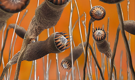
A forest of brown tree-like organisms, set against an orange sky. A vision of Mars, a scene from Star Trek? No, it's a picture of tiny moss capsules by Martin Oeggerli.
Using a highly sophisticated microscope, the Basel scientist takes pictures of the extremely small – a cell, a grain of pollen – and turns them into award-winning scientific fine art.
By day 34-year-old Oeggerli is a molecular biologist working in cancer research at Basel University Hospital. By night, the micronaut, as he likes to be known, heads into the world of colour Scanning Electron Microscope (SEM) photography.
“It’s like you’re flying over a subject,” Oeggerli said of the microscope, which allows him to home in on the tiniest detail. The powerful SEM can magnify a subject up to 500,000 times.
“And it’s the opposite of an astronaut who goes into space because you go into the microcosmos,” he told swissinfo.
The whole process is a mix of science, photography and art. Oeggerli first selects a specimen and prepares it. He then generates a photograph using the SEM.
Afterwards he painstakingly colours the image electronically to his own design, using a special, self-taught, technique. A picture can take up to 100 hours to finish. This takes plenty of patience and a good eye, says Oeggerli.
The moss capsules were captured just as they were breaking open after a dry period to release their spores.
“I tried to design the picture such that if you look at it, you think you are in a moss forest,” said Oeggerli.
“When you really walk through a forest your eyes and brain put images together and you quickly get a 3D impression, and that’s what I tried to imitate,” he explained.
Colouring the microcosmos
Oeggerli tries to be faithful to the real colours of his subjects, which he researches beforehand.
But sometimes, because he is using very high magnification, items become almost transparent, “and there I have my freedom as an artist and can choose colours that fit,” he says.
In reality the moss forest was brown, says Oeggerli, but the orange background and tiny blue spores are his own design.
Sometimes his work turns up surprises, as happened with the butterfly wing helpfully provided by Oeggerli’s cat Herbert.
Magnified 5,300 times the wing reveals scales, coloured blue in the picture, and under them small structures in yellow (see gallery). Ever the curious biologist, Oeggerli tried to find out what they were and consulted an expert.
“It’s not clear what they are for, even for the expert. Maybe they are for pheromone distribution or they could also have an aerodynamic function,” Oeggerli explained.
Alien spores
The biologist-artist looks for his inspiration everywhere. His latest award-winning picture comes from a plant found just outside his door.
This image beat 700 international entries to win first place in the 2008 Best Scientific Cover category at the prestigious EMBO journal of the European Molecular Biology Organisation.
“I found this plant which was infected with little orange dots. I took one of the leaves because I thought it would be a perfect subject to get some more photography practice and to find out what the dots were,” Oeggerli said.
“They were fruit bodies and inside these are hundreds and thousands of fungi spores, which look like little aliens.”
But Oeggerli does not just limit himself to the world of nature, he also gains inspiration from his scientific work and is planning to look at cancer cells.
The amateur photographer knew about the SEM from his studies. But it was not until he was asked to do some artwork for a company annual report that the micronaut’s career took off.
However, finding a high-powered microscope was a problem, as SEMs are very specialised and expensive. Oeggerli solved this by making use of one at a scientific company in canton Uri, in inner Switzerland, in return for his biological expertise.
His lab in Basel, where he works 75 per cent, kindly allows him to use its equipment to prepare specimens.
Scientist or artist?
For now, the micronaut, who has a PhD in biology, has no thoughts of entirely devoting himself to his artwork.
“Science is also very important because from there I also get a lot of ideas… so at the moment it’s fine for me to live in both worlds,” he told swissinfo.
Future projects include taking a close look at butterfly eggs, as well as plant surfaces.
For the passionate explorer of the microscopic world, it is truly fascinating to be able to show people the flipside of the familiar, like a fly’s eye.
“I also want to raise awareness among the public for these beautiful little things which surround us in the microcosmos,” he said.
“Nowadays nature is in danger, so it’s important that we see there are a lot of amazing things and that it’s worth taking care of nature.”
swissinfo, Isobel Leybold-Johnson in Basel
A selection of the micronaut’s work is on display at the Restaurant Union in Basel until the end of the month. Other pictures can be viewed on his site.
Oeggerli is supported in his endeavours by the Institute of Pathology, Basel, Prüftechnik Uri (PTU) and the University of Applied Sciences Northwestern Switzerland: Life Sciences Department.
Apart from the 2008 Best Scientific Cover category at the EMBO journal of cell biology, Oeggerli’s image of tulip pollen gained the third most significant scientific picture of the year accolade at the German Bilder der Forschung international competition.
To do this type of artwork, you need a lot of expertise, which is why only a few people are skilled enough to carry it out. Oeggerli says perfectionism and patience are key.
The SEM uses electrons instead of light to create a highly magnified image.
Specimens have to be prepared very carefully so they can withstand the vacuum in the microscope. Those containing water have to be dried in a way to prevent shrivelling.
Specimens also need to conduct electricity so some need a thin layer of conductive material such as gold.
The SEM is a popular research tool due to its advantages of higher magnification, larger depth of focus, greater resolution, and ease of sample observation.

In compliance with the JTI standards
More: SWI swissinfo.ch certified by the Journalism Trust Initiative




You can find an overview of ongoing debates with our journalists here . Please join us!
If you want to start a conversation about a topic raised in this article or want to report factual errors, email us at english@swissinfo.ch.